GRAM STAIN
DISCOVERED BY
Hans Christian Joachim Gram was a Danish physician and bacteriologist who developed in 1884 the most widely used method of staining bacterial cells for microscopic study.
PRINCIPLE OF GRAM STAIN
- Crystal violet (gentian violet) serves as the primary stain, binding to the bacterial cell wall after treatment with a weak solution of iodine which serves as the mordant for binding the dye.
- Some bacterial species, because of the chemical nature of their cell wall, have ability to retain the dye iodine complex even after treatment with an organic decolouriser such as mixture of equal parts of 95% ethyl alcohol & acetone.
- Dye retaining bacteria stain dark-purple while bacteria which are decolorized take the counter stain (safranine) and appear pink.

PROCEDURE OF GRAM STAIN
1. Lay the fixed slide on a staining rack. Cover the smear completely with the gentian violet. Keep it for 1 minute.
2. Pour off the stain and rinse with running tap water.
3. Flood the slide with Gram’s iodine solution and wait for 2-3 minute.
4. Gently drain off the iodine solution and rinse with running tap water.
5. Decolorize rapidly (few seconds) with acetone/alcohol by holding the smear between thumb & forefinger.
6. Wash the slide under running tap water & drain.
7. Pour safranin solution on the slide and keep for 30-60 seconds.
8. Wash off the stain with tap water.
9. Wipe the back of the slide clean, and place in a draining rack for the smear to air-dry.
10. Examine the smear microscopically with oil immersion objective.
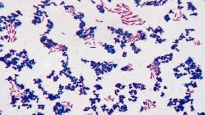
Interpretation of Gram Stain
Gram positive bacteria - Dark purple
Yeast cells - Dark purple
Gram negative bacteria - Pale to dark red
Nuclei of pus cells - Red
Epithelial cells - Pale red
MECHANISM OR THEORIES OF GRAM STAIN
1. Lipid content theory -
- Gram positive organisms contain more protein while gram negative contain more lipid.
- While decolorizing, the lipid get dissolved, thus leaking of dye-iodine complex out of the cell occurs.
- Thus gram negative take the counter stain. In gram positive organisms, the protein in cell wall gets dehydrated, shrinking the pore size.
- Thus do not get decolorized & take primary stain.
2.Magnesium ribonucleate theory –
- Present in Gram Positive Bacteria and there is formation of magnesium ribonucleate – dye – iodine complex which is insoluble in alcohol used as a decolorizer.
3. pH theory –
- As compared to Gram Negative Bacteria, Gram Positive Bacteria have more acidic protoplasm and the primary stain used is basic in nature.
- Hence, Gram Positive Bacteria retain primary stain more strongly than Gram Negative Bacteria.
- Also the iodine makes the cytoplasm more acidic and acts as a mordant, fixes the stain in bacterial cell.
4.Cell wall permeability –
- The cell wall of Gram Positive Bacteria contain more mucopeptide because of which it is thicker & stronger, hence dye iodine complex does not come out of cell easily.
USES OF GRAM STAIN
- Gram stain is useful for presumptive diagnosis of:
- Meningitis from CSF sample - pneumococci, streptococci, meningococci, haemophilus etc.
- Diphtheria from throat swab – by demonstration of Chinese letter pattern of corynebacterium diphtheriae with clubbed ends.
- Upper respiratory tract infection from sputum specimens
- Urinary tract infection
- Dysentery
- Cellulites, osteomyelitis, abscess etc.
- Gas gangrene
- Opportunistic diarrhea in patients on prolonged antibiotic therapy, and various other conditions.
Modifications
of Gram Staining
There are few minor modifications of Gram stain which vary slightly from the method described earlier.
- Kopeloff and Beerman's modification: Primary stain and counter stain used are methyl violet and basic fuchsin respectively.
- Jensen's modification: This method involves use of absolute alcohol as decolorizer and neutral red as counter stain. It is useful for meningococci and gonococci.
- Weigert's modification: This modification is useful for staining tissue sections. Here, aniline-xylol is used as a decolorizer.
- Preston and Morrell's modification: Here, iodine-acetone is used as decolorizer.
NOTE ON GRAM STAIN:
- Different primary stains that can be used in gram stain are methyl violet, gentian violet and crystal violet.
- Different counter stains that can be used in gram stain are safranine, neutral red, dilute carbol fuchsin and basic fuchsin.
- Different decolourisers that can be used in gram stain are acetone, alcohol or a mixture of acetone and alcohol in 1:1 ratio.
- Gram’s iodine acts as a mordant in gram stain. A mordant is a substance which helps in fixation and/or penetration of the dye inside the cell.

No comments:
Post a Comment