Bacterial cell wall is a tough and rigid structure, surrounding the bacterium. It is 10-25 nm in thickness and weighs about 20-25% of the dry weight of the cell.
- Provides protection to the cell against osmotic lysis.
- Confers rigidity upon bacteria due to presence of peptidoglycan layer in the cell wall.
- Cell wall accounts for the shape of the cell.
- Takes part in cell division.
- The cell wall can protect a cell from toxic substances and is the site of action of several antibiotics.
- Virulence factors-Bacterial cell wall contains certain virulence factors (e.g. endotoxin), which contributes to their pathogenicity.
- Immunity: Antibody raised against specific cell wall antigens (e.g. antibody to LPS) may provide immunity against some bacterial infection.
Gram-positive Bacterial Cell Wall
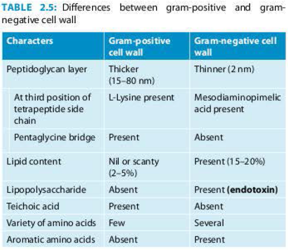
Cell wall of gram-positive bacteria is simpler than that of gram-negative bacteria (Table 2.5).
Peptidoglycan
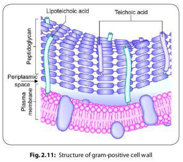
- In gram-positive bacteria, the peptidoglycan layer is much thicker(50- 100 layers thick, 16-80 nm) than gram-negative cell wall (Fig. 2.11).
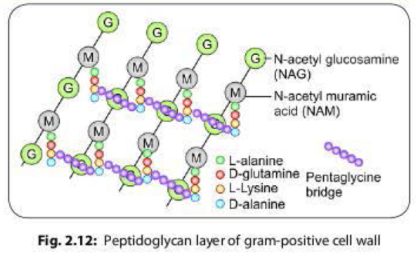
- Each layer is a mucopeptide (murein) chain, composed of alternate units of N-acetyl muramic acid (NAM) and N-acetyl glucosamine (NAG) molecules; cross linked to each other via tetrapeptide side chains and pentaglycine bridges.
- A tetrapeptide side chain ascended from NAM molecule is composed of L-alanine-D-glutamine-L-lysine-D-alanine.
- The L-lysine of one tetrapeplide chain is covalently linked to the terminal D-alanine of the adjacent chain via a pentaglycine bridge. (Fig. 2.12).
Teichoic Acid
- Gram-positive cell wall contains significant amount of teichoic acid which is absent in gram-negative bacteria.
- They are polymers of glycerol or ribitol joined by phosphate groups. The functions of these molecules are still unclear, but they may be important in maintaining the structure of the cell wall. Teichoic acids are of two types:
1. Cell wall teichoic acid: It is covalently linked to NAM molecules of peptidoglycan.
2. Lipoteichoic acid: It is attached to lipid
groups of cell membrane.
Gram-negative Bacterial Cell Wall
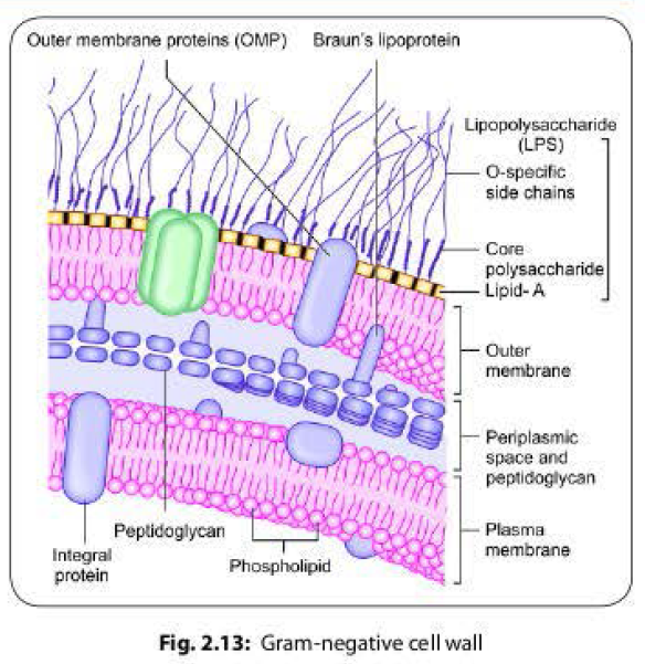
Gram-negative cell wall is thinner and more
complex than the Gram-positive cell wall,
comprises of the following components (Fig.
2.13) (Table 2.5).
Peptidoglycan Layer
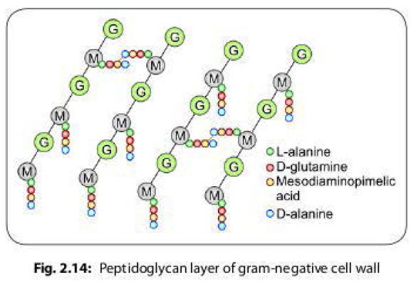
- It is very thin (1-2 layer, 2 nm thick), composed of a mucopeptide chain similar to that of gram-positive cell wall, and consists of alternate NAM and NAG molecules.
- However, it differs from the later by (Fig. 2.14 )-
- Meso-diaminopimelic acid is present at third position of the tetrapeptide side chain ascended form NAM molecule (side chain is composed of L-alanine- D-glutamine-D-mesodiaminopimelic acid-D-alanine)
- The pentaglycine bridge is absent.
- The tetrapeptide side chains are directly linked to each other by the covalent linkage between D-alanine of one chain with mesodiamiopimelic acid of the adjacent chain.
Outer Membrane of Bacteria
- This is a phospholipid layer which lies outside to the thin peptidoglycan layer; firmly attached to the layer by covalent linkage of membrane protein called Braun's lipoprotein.
- It serves as a protective barrier to the cell.
- Outer membrane proteins (OMP) or porin proteins.
- They are the specialized proteins present in outer membrane. Three porin molecules cluster together and span the outer membrane to form a narrow channel through which molecules smaller than about 600-700 daltons can pass.
- Larger molecules such as vitamin B must be transported across the outer membrane by specific carriers.
- The outer membrane also prevents the loss of constituents such as periplasmic enzymes.
Lipopolysaccharide (LPS)
This layer is unique to gram-negative bacteria which is absent in gram-positives. It consists of three parts:
Lipid A or the Endotoxin:
- It has endotoxic activities, such as pyrogenicity, lethal effect, tissue necrosis, anticomplementary activity, B cell mitogenicity, adjuvant property and antitumour activity.
- It consists of two glucosamine sugar derivatives, each with three fatty acids and phosphate attached. lt is buried in the outer membrane and the remainder of the LPS molecule projects from the surface.
Core polysaccharide:
- It is projected from lipid A region. lt is composed of 10- 12 sugar moieties.
O side chain (or O antigen):
- It is a polysaccharide chain extending outwards from the core polysaccharide region. It is made up of several sugar moieties and it greatly varies in composition between bacterial strains.
- O antigen is a major surface antigen(called somatic antigen), induces antibody formation. It is also used for serotyping.
Periplasmic Space
- It is the space between the inner cell membrane and outer membrane. It encompasses the peptidoglycan layer.
Demonstration
of the Bacterial Cell Wall
The cell wall cannot be seen by light microscope and does not stain with simple dyes. Demonstration of cell wall can be done by methods such as:
- Plasmolysis: When bacteria are placed in a hypertonic saline, shrinkage of the cytoplasm occurs, while cell wall retains original shape and size.
- Microdissection
- Differential staining
- Reaction with specific antibody
- Electron microscopy

No comments:
Post a Comment