There are two types of variations seen in bacteria:
- Phenotypic variation in bacteria: It refers to the variations in the expression of various characters by bacterial cells in a given environment, such as synthesis of flagella, expression of certain enzymes, etc.
- Genotypic variation in bacteria: It is the change in the genetic constitution of an organism; which occurs mostly as a result of mutation.
MUTATION IN BACTERIA
Definition: Bacterial mutation
is a random, undirected heritable variation caused by change in nucleotide
sequence of the genome of the cell.
Bacterial mutation can
involve any of the numerous genes present in bacterial chromosome or rarely
plasmid. The frequency of mutation ranges from 10-2 to 10-10
per bacterium per division.
Bacterial mutations
occur in one of the two ways:
1. Spontaneous mutations in bacteria: Mutations that occur naturally in any dividing cells that arise occasionally without adding any mutagen.
2. Induced mutations in bacteria: : These mutations on the other hand, are as a result of exposure of the organism to a mutagen, an agent capable of inducing mutagenesis.
Examples of mutagens include-
Physical agent, e.g. ultraviolet (UV) radiations, cytosine and thymine are more vulnerable to UV rays.
Chemical agents, e.g. alkylating agents, 5-bromouracil and acridine dyes.
Bacterial mutation is a
natural event, taking place all the time, in all dividing cells. Most mutants
go unrecognized as the mutation may be lethal or may involve some minor functions
that may not be expressed. Bacterial mutation is best appreciated when it involves a function,
which can be readily observed by experimental methods. For example E.coli mutant that loses its ability
to ferment lactose can be readily detected on MacConkey agar.
Mutation can
affect any gene and hence may modify any characteristic of the bacterium, for
example:
- Sensitivity to bacteriophages
- Loss of ability to produce capsule or flagella
- Loss of virulence
- Alteration in colony morphology
- Alteration in pigment production
- Drug susceptibility
- Biochemical reactions
- Antigenic structure
The practical
importance of bacterial mutation is mainly in the field of drug resistance and
the development of live vaccines.
Classification
of Bacterial Mutation Types
Mutations may
occur in two ways-
1. Small-scale mutations: They are more commonly seen in bacteria. Examples include (1) point mutations- occur at a single nucleotide, (2) addition or deletion of single nucleotide pair
2. Large-scale mutations: Occur in chromosomal structure. These include deletion or addition of several nucleotide base pairs or gene duplications.
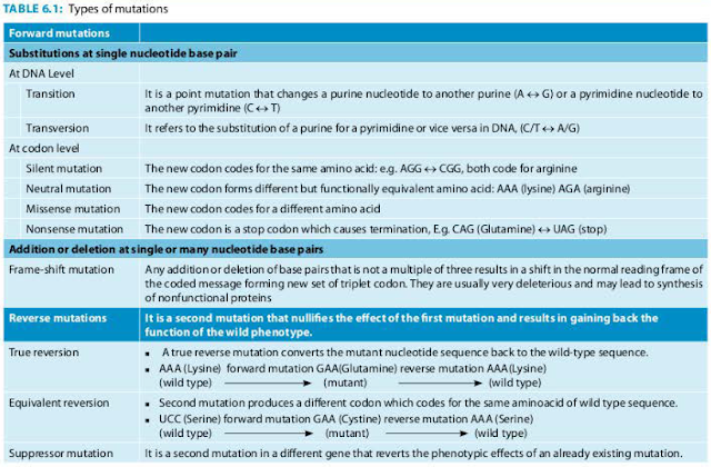
Various types of mutations observed in bacteria are described in Table 6. 1.
Detection and Isolation
of Bacterial Mutants
Mutation can
be recognized both by genetic method (gene sequencing) as well as by observing
phenotypic changes such as fluctuation test and replica plating method. Like carcinogenicity
of a mutagen is tested by Ames lest.
Fluctuation Test to Detect Bacterial Mutation
Fluctuation
test demonstrates the spontaneous mutation in bacteria. It was described by Luria
and Delbruck (1943).
- It states that when bacterial suspension is subjected to selective pressure by sub-culturing on to agar plate containing a growth limiting substance (e.g. streptomycin or bacteriophage, etc.), they undergo spontaneous mutation.
- However, the rate of mutation vary widely (some bacteria mutate early, some late) which leads to fluctuations.
- Fluctuations in mutation are wide when small volume sub cultures are made (which leads to more frequent mutations), as compared to large volume subcultures (where the mutations occur less frequently).
- This experiment was not widely appreciated, probably due to the complicated statistical evaluation.
Replica Plating Method
Replica plating
method is used to detect auxotrophic mutations, described by Lederberg in 1952.
It differentiates between the normal strains from auxotrophic mutants based on
their ability to grow in the absence of a particular nutrient on which the
mutant is dependent. For example, a lysine auxotroph will grow on
lysine-supplemented media but not on a medium lacking lysine.
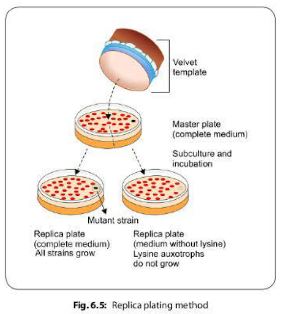
- Using a velvet template, mixture of colonies (some normal strain, some auxotroph mutants) of an organism are transferred from a master plate, onto two subculture plates--One of the plate is lacking a limiting nutritional substance, e.g. lysine.
- After incubation, colonies similar to those on master plate are formed with
relative position of all the colonies retained
on the subculture plates, except for the lysine auxotroph which do not grow on the media lacking lysine (Fig. 6.5).
Ames Test (Carcinogenicity Testing)
Ames test is used
to identify the environmental carcinogens.
It was developed
by Bruce Ames (1970).
- It is a mutational reversion assay that uses the mutant strains (histidine auxotroph) of Salmonella which are sub-cultured on two agar plates containing small amount of histidine; one of the plate is added with the test mutagen.
- The plates are incubated for 2-3 days at 37°C.
- All of the histidine auxotrophs will grow for the first few hours until the histidine is depleted.
- Once the histidine supply is exhausted, only revertants that have mutationally regained the ability to synthesize histidine will grow (Fig. 6.6).
- Reversed mutation may be induced due to carcinogen (can affect large number of strains) or occur spontaneously (affect only few strains).
- The relative mutagenicity of the carcinogen can be estimated by counting the colonies-the more colonies, the greater is the mutagenicity.


No comments:
Post a Comment