Fluorescence
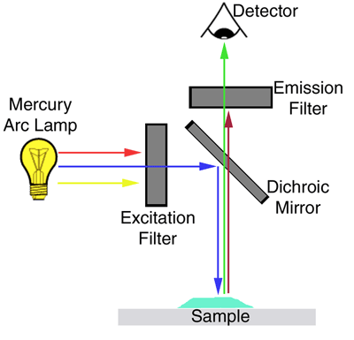
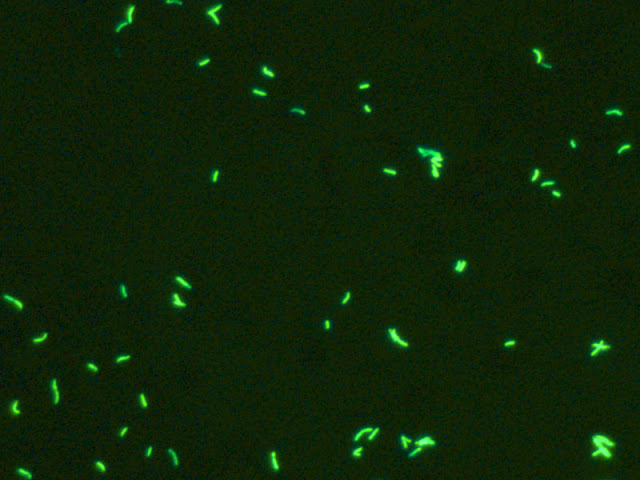
microscope is based on the principle of removal of incident
illumination by selective absorption, whereas light that has been absorbed by
the specimen and re-emitted at an altered wavelength is transmitted.
The light
source must produce a light beam of appropriate wavelength. An excitation
filter removes wavelengths that are not effective in exciting the fluorochrome
used.
The light fluoresced by the specimen is transmitted through a filter that
removes the incident wavelength from the beam of light. As a result, only light
that has been produced by specimen fluorescence contributes to the intensity of
the image being viewed.
- Acridine orange
- Rhodamine-auramine
- Calcofluor white
- Fluorescein isothiocyanate
- Lissamine rhodamine
USES OF FLUORESCENCE MICROSCOPE:
1. For staining of tissues, cells or
bacteria by using dyes like acridine orange, auramine O, calcofluor white, etc.
2. For immunofluorescence by using fluorescent dyes like fluorescein
isothiocyanate & lissamine rhodamine to label the antibodies or antigens.


No comments:
Post a Comment